toh
Interventional Radiology
What is an Interventional Radiology?
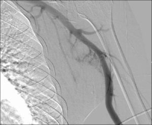
Interventional Radiology (IR) also known as Angiography.
Interventional radiology (IR) is a medical specialty that performs minimally invasive treatments using x ray imaging for procedure guidance. Interventional radiologists use x-ray imaging to guide small instruments, like catheters, through blood vessels and organs to treat a variety of diseases.
How does it work?
- IR procedures use imaging equipment to assist a physician in the treatment of a patient’s condition.
- Most IR procedures begin with a small prick of a needle in either in the arm or groin area. Then your interventional radiologist—who is trained in radiology and minimally invasive interventional therapies—guides a thin wire and a catheter, through a blood vessel to reach and treat the source of your pain or disease.
What is it used for?
An X-ray exam of the arteries and veins to diagnose blockages and other blood vessel problems; uses a catheter to enter the blood vessel and a contrast agent (X-ray dye) to make the artery or vein visible on the X-ray.
Some examples of angiography procedures include angioplasty, stenting, thrombolysis, embolization, radiofrequency ablation, and biopsies. These procedures can treat or help symptoms of vascular disease, stroke, uterine fibroids, or cancer.

Safety information
With interventional radiology procedures using x-rays, the level of risk depends on the type of procedure because some use very little radiation, while complex procedures use much more.
You need to let us know if you are pregnant or have any serious allergy reactions.
Angiography is a very safe procedure, but as with any medical procedure, there are some risks and complications that can arise. These will be discussed with you on the day of the procedure or in a pre procedure clinic visit.
Always talk with your doctor to understand the risks and benefits.
Intravascular (IV) contrast agent
Intravascular contrast agent is an iodine containing solution. Because it shows up white on X-ray images, it is sometimes referred to as “X-ray dye”. Your doctor has requested and recommends an examination that requires injection of contrast agent either into a vein or artery. This injection will enable the Radiologist to better see specific areas of your body. At TOH we only use a non-ionic contrast agents for your safety.
How to prepare?
Before your procedure
- Ask all the questions you want.
- Be sure you understand any pre-procedure tests (e.g. bloodwork), medications, special precautions, transportation and necessary preparations. Please do not wear jewelry or bring valuables with you.
- Location of your appointment will be provided by the booking clerk prior to your arrival.
- Do not eat or drink after midnight before procedure, unless otherwise directed.
- Morning of procedure, take blood pressure and/or heart medications with a sip of water unless told otherwise.
- If you are diabetic, take you insulin and a small meal unless otherwise directed.
- You will not be allowed to drive after your exam, so please have transportation home arranged.
- Ask your doctor about discontinuing aspirin or blood thinners seven days before procedure.
- Tell the staff if you have NOT had lab work or are on any medication.
- If you are pregnant or think you might be, tell the staff before the test.
- If you have any allergies to medications, or X-ray dye, tell the staff before the test.
On day of your procedure
The day of your procedure, you will arrive as instructed and a team of experts will prepare you and answer any remaining questions.
Most IR procedures are done using a local freezing and. you’ll be relaxed, but awake during the entire procedure. Some procedures may require that you receive a medicine that puts you to sleep (general anesthesia). You will be taken care of by the IR team of nurses, medical radiation technologists and interventional radiologists, who will keep you comfortable during your treatment. Your care team will also take precautions to ensure complete safety.
After your procedure
Once your procedure is completed, you will be moved to the recovery area. Depending on your procedure, you may be discharged the same day or following a short hospital stay. Your care team will review your recovery and discharge time, restrictions when you go home, possible complications and necessary medications.
You will receive information on follow up care.
After you go home
The contrast agent is gradually removed from your blood by your kidneys as you urinate. The colour of your urine will not change . You may immediately resume your normal diet. You are encouraged to drink several glasses of water (as tolerated or dependent on any fluid restrictions you may have)within the next 24 hours to help your body remove the contrast agent
Your injection/needle puncture site should be watched for swelling or bleeding. If it remains dry, you may remove the bandage or dressing, unless otherwise indicated in post care instructions
Call your doctor for advice if:
- Significant swelling or bleeding occurs at the injection/puncture site
- Your experience any hives or itching
- You experience any difficulty in swallowing or breathing
- IF you cannot get in touch with your own doctor, go to the closest Emergency department
What type of exams are performed in Interventional Radiology?
Balloon angioplasty
Opens blocked or narrowed blood vessels by inserting a very small balloon into the vessel and inflating it. Used to unblock clogged arteries in the legs or arms (called peripheral vascular disease or PVD), kidneys, brain or elsewhere in the body.
Biopsies
A needle is used to collect a tissue sample from somewhere in your body e.g the Lung, Bone, Liver: use of needles to obtain cell samples, an alternative to surgical biopsy.
Central venous access
Placement of a tube beneath the skin and into the blood vessels so that patients can receive medication or nutrients directly into the blood stream or so blood can be drawn. (PORT, Hickman lines, PICC)
Chemoembolization
Delivery of cancer-fighting agents (alcohol, chemo.) directly to the site of a cancer tumor; currently being used mostly to treat cancers.
Embolization
Delivery of clotting agents (coils, plastic particles, gelfoam, etc.) directly to an area that is bleeding or to block blood flow to a problem area, such as an aneurysm, Aterial venous malformation (AVM) or a fibroid tumor in the uterus.
Gastrostomy tube Insertion
Feeding tube inserted into the stomach for patients who are unable to take sufficient food by mouth.
Hemodialysis and access maintenance
insertion of catheters and follow up use of angioplasty or thrombolysis to open blockages for hemodialysis.
IVC filter
Placement/retrieval of temporary or permanent filters in the Inferior Vena Cava to prevent blood clots moving into the lungs.
Radiofrequency (RF) ablation
Use of radiofrequency (RF) energy to destroy cancerous tumors.
Spinal procedures
Vertebroplasty, Myelograms, Facet injections, epidural injections, Discograms, lumbar puncture, Nerve blocks, Rhizotomy – various procedure for spinal problems
Stent
A small flexible tube made of plastic or wire mesh, used to treat a variety of medical conditions (e.g., to hold open clogged blood vessels or other pathways that have been narrowed or blocked by tumors or obstructions). Some examples of stents are Carotid, iliac, femoral, SVC, biliary duct.
Stroke
Strokes caused by blood clots can be dissolved (thrombolysis) or by removal (mechanical thrombectomy). Strokes caused by bleeding resulting from ruptured aneurysms may be treated by embolization, most commonly using tiny metal coils.
Thrombolysis
Dissolves blood clots by injecting clot-busting drugs at the site of the clot.
TIPS (transjugular intrahepatic portosystemic shunt)
The insertion of a devise that is used to improve blood flow and prevent bleeding in patients with severe liver problems.
Miscellaneous
Removal of fluid (Drainage), and the Injection of medicine into a body part.
Last updated on: March 12th, 2021


 To reset, hold the Ctrl key, then press 0.